Articles
-
What ails the NIH peer review study sections and how to fix the review process of the grant applications
J Cardiovasc Aging 2023;3:11. DOI: 10.20517/jca.2023.3Perspective|Published on: 16 Jan 2023 -
Parsing cell death in arrhythmogenic cardiomyopathy: PANoptosis
J Cardiovasc Aging 2023;3:10. DOI: 10.20517/jca.2022.45Editorial|Published on: 12 Jan 2023 -
The dynamic interplay between cardiac mitochondrial health and myocardial structural remodeling in metabolic heart disease, aging, and heart failure
J Cardiovasc Aging 2023;3:9. DOI: 10.20517/jca.2022.42AbstractThis review provides a holistic perspective on the bi-directional relationship between cardiac mitochondrial dysfunction and ... MOREThis review provides a holistic perspective on the bi-directional relationship between cardiac mitochondrial dysfunction and myocardial structural remodeling in the context of metabolic heart disease, natural cardiac aging, and heart failure. First, a review of the physiologic and molecular drivers of cardiac mitochondrial dysfunction across a range of increasingly prevalent conditions such as metabolic syndrome and cardiac aging is presented, followed by a general review of the mechanisms of mitochondrial quality control (QC) in the heart. Several important mechanisms by which cardiac mitochondrial dysfunction triggers or contributes to structural remodeling of the heart are discussed: accumulated metabolic byproducts, oxidative damage, impaired mitochondrial QC, and mitochondrial-mediated cell death identified as substantial mechanistic contributors to cardiac structural remodeling such as hypertrophy and myocardial fibrosis. Subsequently, the less studied but nevertheless important reverse relationship is explored: the mechanisms by which cardiac structural remodeling feeds back to further alter mitochondrial bioenergetic function. We then provide a condensed pathogenesis of several increasingly important clinical conditions in which these relationships are central: diabetic cardiomyopathy, age-associated declines in cardiac function, and the progression to heart failure, with or without preserved ejection fraction. Finally, we identify promising therapeutic opportunities targeting mitochondrial function in these conditions. LESS Full articleReview|Published on: 3 Jan 2023 -
Lipid overload - a culprit for hypertrophic cardiomyopathy?
J Cardiovasc Aging 2023;3:8. DOI: 10.20517/jca.2022.43Editorial|Published on: 1 Jan 2023 -
Thrombosis and myocardial infarction: the role of bioresorbable scaffolds
J Cardiovasc Aging 2023;3:7. DOI: 10.20517/jca.2022.41AbstractCoronary atherosclerosis is a leading cause of death as a result of coronary thrombosis and ... MORECoronary atherosclerosis is a leading cause of death as a result of coronary thrombosis and acute myocardial infarction. Drug-eluting stents (DES) have dramatically improved the treatment of coronary artery stenosis. However, stent thrombosis (ST) and in-stent-restenosis (ISR) have remained a vexing limitation of the DES. After DES implantation, despite taking dual antiplatelet (DAPT) therapy, very late ST results in myocardial infarction and death. This occurs regardless of the type of polymer or antiproliferative agent used in the contemporary DES. Such adverse events occur at a rate of approximately 2% to 3% per year after the first year, which have been attributed to strut fractures, loss of vessel compliance, and neoatherosclerosis. Bioresorbable scaffolds (BRS) have been introduced to overcome the above shortfalls and to a “leave nothing behind” approach. While BRS are novel and interesting, the initial experience with BRS was hampered by the increased rate of thrombosis compared with DES. Accordingly, in this review, we summarized underlying mechanisms leading to BRS failure and provided insights into optimizing BRS deployment with intravascular imaging. In addition, we outlined the perspectives of new generations of BRS with thinner struts and new designs as well as alternative materials to improve outcomes. LESS Full articleReview|Published on: 1 Jan 2023 -
The TNNI3 p.R186Q mutation is responsible for hypertrophic cardiomyopathy via promoting FASN-stimulated abnormal fatty acid metabolism
J Cardiovasc Aging 2023;3:6. DOI: 10.20517/jca.2022.29AbstractIntroduction: The TNNI3 gene encodes the protein of cardiac troponin I (cTnI), which is an ... MOREIntroduction: The TNNI3 gene encodes the protein of cardiac troponin I (cTnI), which is an inhibitory subunit of sarcomeres. Mutations in this gene account for 3% of hypertrophic cardiomyopathy (HCM) and the molecular mechanism is complex. Recently, lipid metabolism has been revealed to be involved in HCM. Aim: The purpose of this work is to identify whether the pathological mechanism of the hotspot mutation TNNI3 p.R186Q in HCM is related to abnormal lipid metabolism.Methods and Results: A knock-in (KI) mouse model carrying the Tnni3 p.R186Q homozygous mutation(Tnni3R186Q/R186Q) was novelty generated by CRISPR/Cas9 technology and successfully constructed a typical phenotype of cardiac-myopathy. Likewise, neonatal rat cardiomyocytes (NRCMs) transfected with a mutant plasmid with the TNNI3 p.R186Q mutation showed the same phenomenon. In-depth experiments on related functions and molecular mechanisms were conducted, and Tnni3R186Q/R186Q mice exhibited abnormal fatty acid metabolism, which was induced by the activation of epidermal growth factor receptor(EGFR)-dependent high expression of fatty acid synthase (FASN) in vivo and in vitro. Specifically, the direct binding of EGFR and cTnI was destroyed by TNNI3 p.R186Q mutation, as observed through bioinformatics, Co-IP and GST-pull down analysis.Conclusion: In the present study, we successfully engineered Tnni3R186Q/R186Q mice with the typical phenotype of myocardial hypertrophy. We demonstrated that the TNNI3 p.R186Q mutation could induce HCM by the dissociation of EGFR and cTnI, which further led to EGFR-dependent increased expression of FASN and abnormal lipid metabolism. LESS Full articleOriginal Research Article|Published on: 1 Jan 2023 -
The importance of “when” in calorie restriction-induced lifespan extension
J Cardiovasc Aging 2023;3:5. DOI: 10.20517/jca.2022.40Commentary|Published on: 1 Jan 2023 -
PANoptosis is a prominent feature of desmoplakin cardiomyopathy
J Cardiovasc Aging 2023;3:3. DOI: 10.20517/jca.2022.34AbstractIntroduction: Arrhythmogenic cardiomyopathy (ACM) is hereditary cardiomyopathy caused by pathogenic variants (mutations) in genes encoding ... MOREIntroduction: Arrhythmogenic cardiomyopathy (ACM) is hereditary cardiomyopathy caused by pathogenic variants (mutations) in genes encoding the intercalated disc (ID), particularly desmosome proteins. ACM caused by mutations in the DSP gene encoding desmoplakin (DSP) is characterized by the prominence of cell death, myocardial fibrosis, and inflammation, and is referred to as desmoplakin cardiomyopathy.Aim: The aim of this article was to gain insight into the pathogenesis of DSP cardiomyopathy.Methods and Results: The Dsp gene was exclusively deleted in cardiac myocytes using tamoxifen-inducible MerCreMer (Myh6-McmTam) and floxed Dsp (DspF/F) mice (Myh6-McmTam:DspF/F). Recombination was induced upon subcutaneous injection of tamoxifen (30 mg/kg/d) for 5 days starting post-natal day 14. Survival was analyzed by Kaplan-Meier plots, cardiac function by echocardiography, arrhythmias by rhythm monitoring, and gene expression by RNA-Seq, immunoblotting, and immunofluorescence techniques. Cell death was analyzed by the TUNEL assay and the expression levels of specific markers were by RT-PCR and immunoblotting. Myocardial fibrosis was assessed by picrosirius red staining of the myocardial sections, RT-PCR, and immunoblotting. The Myh6-McmTam:DspF/F mice showed extensive molecular remodeling of the IDs and the differential expression of ~10,000 genes, which predicted activation of KDM5A, IRFs, and NFκB and suppression of PPARGC1A and RB1, among others in the DSP-deficient myocytes. Gene set enrichment analysis predicted activation of the TNFα/NFκB pathway, inflammation, cell death programs, and fibrosis. Analysis of cell death markers indicated PANoptosis, comprised of apoptosis (increased CASP3, CASP8, BAD and reduced BCL2), necroptosis (increased RIPK1, RIPK3, and MLKL), and pyroptosis (increased GSDMD and ASC or PYCARD) in the DSP-deficient myocytes. Transcript levels of the pro-inflammatory and pro-fibrotic genes were increased and myocardial fibrosis comprised ~25% of the myocardium in the DSP-deficient hearts. The Myh6-McmTam:DspF/F mice showed severe cardiac systolic dysfunction and ventricular arrhythmias, and died prematurely with a median survival rate of ~2 months.Conclusion: The findings identify PANoptosis as a prominent phenotypic feature of DSP cardiomyopathy and set the stage for delineating the specific molecular mechanisms involved in its pathogenesis. The model also provides the opportunity to test the effects of pharmacological and genetic interventions on myocardial fibrosis and cell death. One sentence summary: Post-natal homozygous deletion of the Dsp gene leads to fulminant PANoptosis and severe myocardial fibrosis, cardiac dysfunction, arrhythmias, and premature death in mice. LESS Full articleOriginal Research Article|Published on: 1 Jan 2023 -
New insights into the dynamics of age-related clonal hematopoiesis
J Cardiovasc Aging 2023;3:2. DOI: 10.20517/jca.2022.38Commentary|Published on: 1 Jan 2023 -
The role of paracrine crosstalk between myeloid and endothelial cells in myocardial angiogenesis and infarcted heart repair
J Cardiovasc Aging 2023;3:1. DOI: 10.20517/jca.2022.37Commentary|Published on: 1 Jan 2023
See more
Most Downloaded Articles Published In
-
Epigenetic dysregulation in cardiovascular aging and disease
J Cardiovasc Aging 2021;1:10. DOI: 10.20517/jca.2021.16AbstractCardiovascular disease (CVD) is the leading cause of mortality and morbidity for all sexes, racial ... MORECardiovascular disease (CVD) is the leading cause of mortality and morbidity for all sexes, racial and ethnic groups. Age, and its associated physiological and pathological consequences, exacerbate CVD incidence and progression, while modulation of biological age with interventions track with cardiovascular health. Despite the strong link between aging and CVD, surprisingly few studies have directly investigated heart failure and vascular dysfunction in aged models and subjects. Nevertheless, strong correlations have been found between heart disease, atherosclerosis, hypertension, fibrosis, and regeneration efficiency with senescent cell burden and its proinflammatory sequelae. In agreement, senotherapeutics have had success in reducing the detrimental effects in experimental models of cardiovascular aging and disease. Aside from senotherapeutics, cellular reprogramming strategies targeting epigenetic enzymes remain an unexplored yet viable option for reversing or delaying CVD. Epigenetic alterations comprising local and global changes in DNA and histone modifications, transcription factor binding, disorganization of the nuclear lamina, and misfolding of the genome are hallmarks of aging. Limited studies in the aging cardiovascular system of murine models or human patient samples have identified strong correlations between the epigenome, age, and senescence. Here, we compile the findings in published studies linking epigenetic changes to CVD and identify clear themes of epigenetic deregulation during aging. Pending direct investigation of these general mechanisms in aged tissues, this review predicts that future work will establish epigenetic rejuvenation as a potent method to delay CVD. LESS Full articleReview|Published on: 23 Aug 2021 -
Editors’ Preamble to The Journal of Cardiovascular Aging
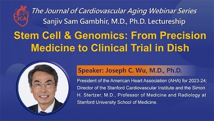
J Cardiovasc Aging 2021;1:1. DOI: 10.20517/jca.2021.01Editorial|Published on: 27 Apr 2021 -
Ser9 phosphorylation of GSK-3β promotes aging in the heart through suppression of autophagy
J Cardiovasc Aging 2021;1:9. DOI: 10.20517/jca.2021.13AbstractIntroduction: Glycogen synthase kinase-3β (GSK-3β) is a serine/threonine kinase and a negative regulator of cardiac ... MOREIntroduction: Glycogen synthase kinase-3β (GSK-3β) is a serine/threonine kinase and a negative regulator of cardiac hypertrophy. Phosphorylation of GSK-3β at Ser9 negatively regulates its kinase activity. The role of GSK-3β in cardiac aging remains poorly understood.Aim: The study aimed to elucidate the role of GSK-3β Ser9 phosphorylation in mediating cardiac aging and the underlying mechanism.Methods and Results: Phosphorylation of GSK-3β at Ser9 and the levels of β-catenin and Mcl-1 were increased in the mouse heart during aging, suggesting that GSK-3β is inactivated during aging in the heart. Age-induced cardiac hypertrophy, fibrosis, left ventricular dysfunction, and increases in cardiomyocyte apoptosis and senescence were all attenuated in constitutively active GSK-3βS9A knock-in (KI) mice compared to littermate wild type mice. Although autophagy is inhibited in the heart during aging, KI of GSK-3βS9A reversed the age-associated decline in autophagy in the mouse heart. GSK-3β directly phosphorylates Ulk1, a regulator of autophagy, at Ser913, thereby stimulating autophagy in cardiomyocytes. Ulk1Ser913A KI mice exhibited decreased autophagic flux and increased senescence in cardiomyocytes.Conclusion: Our results suggest that GSK-3β is inactivated during aging through Ser9 phosphorylation, which in turn plays an important role in mediating cardiac aging. GSK-3β promotes autophagy through phosphorylation of Ulk1 at Ser913, which in turn prevents aging in the heart.One sentence summary: Ser9 phosphorylation of GSK-3β promotes aging in the heart at least in part through decreases in Ulk1 phosphorylation at Ser913 and suppression of autophagy. LESS Full articleOriginal Research Article|Published on: 23 Aug 2021 -
Adenine base editing to treat progeria syndrome and extend the lifespan
J Cardiovasc Aging 2021;1:8. DOI: 10.20517/jca.2021.10Commentary|Published on: 17 Jun 2021 -
S-nitrosoglutathione reductase (GSNOR) deficiency accelerates cardiomyocyte differentiation of induced pluripotent stem cells
J Cardiovasc Aging 2021;1:13. DOI: 10.20517/jca.2021.19AbstractIntroduction: Induced pluripotent stem cells (iPSCs) provide a model of cardiomyocyte (CM) maturation. Nitric oxide ... MOREIntroduction: Induced pluripotent stem cells (iPSCs) provide a model of cardiomyocyte (CM) maturation. Nitric oxide signaling promotes CM differentiation and maturation, although the mechanisms remain controversial.Aim: The study tested the hypothesis that in the absence of S-nitrosoglutathione reductase (GSNOR), a denitrosylase regulating protein S-nitrosylation, the resultant increased S-nitrosylation accelerates the differentiation and maturation of iPSC-derived cardiomyocytes (CMs).Methods and Results: iPSCs derived from mice lacking GSNOR (iPSCGSNOR-/-) matured faster than wildtype iPSCs (iPSCWT) and demonstrated transient increases in expression of murine Snail Family Transcriptional Repressor 1 gene (Snail), murine Snail Family Transcriptional Repressor 2 gene (Slug) and murine Twist Family BHLH Transcription Factor 1 gene (Twist), transcription factors that promote epithelial-to-mesenchymal transition (EMT) and that are regulated by Glycogen Synthase Kinase 3 Beta (GSK3β). Murine Glycogen Synthase Kinase 3 Beta(Gsk3β) gene exhibited much greater S-nitrosylation, but lower expression in iPSCGSNOR-/-. S-nitrosoglutathione (GSNO)-treated iPSCWT and human (h)iPSCs also demonstrated reduced expression of GSK3β. Nkx2.5 expression, a CM marker, was increased in iPSCGSNOR-/- upon directed differentiation toward CMs on Day 4, whereas murine Brachyury (t), Isl1, and GATA Binding Protein(Gata4) mRNA were decreased, compared to iPSCWT, suggesting that GSNOR deficiency promotes CM differentiation beginning immediately following cell adherence to the culture dish-transitioning from mesoderm to cardiac progenitor.Conclusion: Together these findings suggest that increased S-nitrosylation of Gsk3β promotes CM differentiation and maturation from iPSCs. Manipulating the post-translational modification of GSK3β may provide an important translational target and offers new insight into understanding of CM differentiation from pluripotent stem cells.One sentence summary: Deficiency of GSNOR or addition of GSNO accelerates early differentiation and maturation of iPSC-cardiomyocytes. LESS Full articleOriginal Research Article|Published on: 7 Sep 2021
-
Cancer treatment-induced NAD+ depletion in premature senescence and late cardiovascular complications
Review|Published on: 29 Apr 2022 -
Promoting healthy cardiovascular aging: emerging topics
Review|Published on: 29 Jul 2022 -
Metabolic targets in cardiac aging and rejuvenation
Review|Published on: 14 Sep 2022 -
Gut microbiota in sarcopenia and heart failure
Review|Published on: 5 Jul 2022 -
STEMIN and YAP5SA synthetic modified mRNAs regenerate and repair infarcted mouse hearts
Original Research Article|Published on: 15 Jun 2022
See more
About The Journal
-
ISSN
2768-5993 (Online)
Publisher
OAE Publishing Inc.
Article Processing Charges
$1200
Manuscript Processing Cycle (peer reviewed only)
Time from submission to first decision: 18 days (4-38 days)
Time from submission to publication: 31 days (12-48 days)
(not include the time that authors spend on revision)
-
Editor-in-Chief
Ali J. Marian
Publishing Model
Gold Open Access
Copyright
Copyright is retained by author(s)
-
Publication Frequency
Quarterly
Indexing
Open Archives
-
Portico
All published articles are preserved here permanently:
https://www.portico.org/publishers/oae/