
Exercise induces cardiomyogenesis in the aged heart
 0
0
Adult mammalian cardiomyocytes have a limited, but measurable, capacity for self-renewal, which decreases with age. A recent study demonstrates that exercise, in part, restores cardiomyogenesis in aged mice through pathways associated with circadian rhythm[1].
Although aging is regarded as a natural process and most experts do not classify it as a disease, it is an undeniable risk factor for developing certain diseases. As such, aging fits the given medical definition of a disease where “harmful abnormality of bodily structure and function” ensues. Moreover, differential transcriptome analysis of young and aging mouse hearts has revealed changes in several pathways such as hypertrophy, inflammation, and metabolism, which may contribute to the phenotypic changes seen in the aging heart. Importantly, these changes in many ways mimic those seen in the failing heart[2].
The Richard and the Rosenzweig groups showed in 2018 that voluntary exercise for 8 weeks increased cardiomyogenesis in adult mice by up to ~4.6-fold. They demonstrated in the same study that exercise induces a cardiomyogenic response in the myocardial infarction border zone, which supports a pro-regenerative role of exercise. Mechanistically, they showed that miR-222 is necessary for exercise-induced cardiomyogenesis[3]. Studies by other groups also showed that exercise may promote cardiomyocyte proliferation through the induction of growth factors, cell cycle regulators, and transcriptional factors[4].
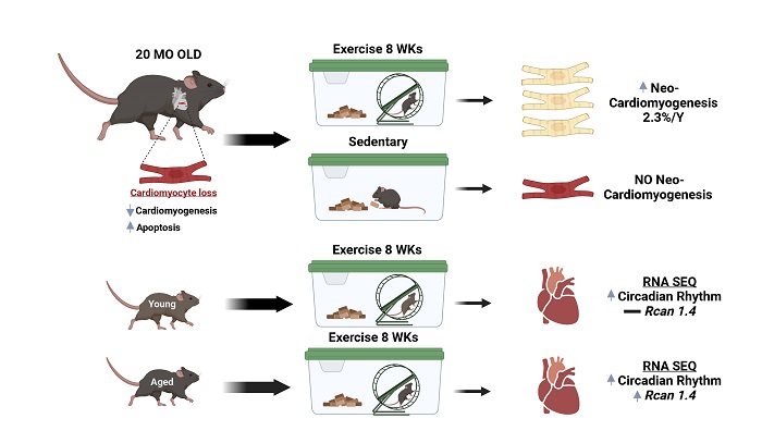
In a recent report in Circulation, the Lee group led by Lerchenmüller et al. expanded their earlier work to the aging hearts in two noteworthy directions[1]. First, they investigated whether exercise also induces cardiomyogenesis in aged hearts. They used MIMS tracking (which the group has previously pioneered) to quantify the integration of 15N-thymidine in cardiomyocyte nuclei in aged mouse hearts after 8 weeks of voluntary running or sedentary activity. Furthermore, they quantified both nucleation and ploidy to show that exercised aged mice generate new mononucleated, diploid, and 15N-thymidine-positive cardiomyocytes with a calculated annual rate of 2.3%, which was not found in the sedentary group. Additionally, the author explains that although the total number of new cardiomyocytes in exercised aged hearts is less than in exercised young hearts (0.35% in exercised aged mice compared with 1.15% in exercised young mice), the relative increase in exercise-induced cardiomyogenesis in aged animals is no less than in young animals[3].
Next, the authors sought to define pathways that mediate exercise-induced cardiomyogenesis in the aged heart. This was achieved by performing RNA seq using whole heart samples from exercised young and aged mice and their age-matched sedentary controls, followed by bioinformatic analysis. Interestingly, they found that both the young and aged exercised hearts were enriched in pathways related to circadian rhythm. However, one gene, Rcan1.4, but not Rcan1.1, was uniquely upregulated in aged, exercised hearts compared with young, exercised hearts, which was further confirmed by QPCR and WB analysis using isolated cardiomyocytes from both groups. This finding is important not only because Rcan1 expression is known to fluctuate in a circadian manner in healthy hearts[5] but more importantly because it was previously shown to be involved in cardiomyocyte proliferation[6], where calcineurin was shown to regulate the growth mode switch of postnatal cardiomyocytes.
Given the known role of Rcan1 in regulation of calcineurin, the authors further investigated the effects of the exercise-induced Rcan1 on calcineurin activity, where they observed reduced NFATc1 nuclear immunostaining in aged, exercised hearts compared to aged sedentary hearts, indicating reduced calcineurin activity with exercise. To further endorse their proliferative phenotype in exercised aged hearts, QPCR results showed the cell cycle promoting gene Cdc2a and its downstream target Ccnd1 mRNA to be upregulated in aged, exercised hearts compared to age sedentary hearts. Furthermore, both Cdc2a and Ccnd1 expression were increased specifically by adenoviral Rcan1.4 expression, rather than Rcan1.1, in isolated aged murine cardiomyocytes, suggesting a proliferative role of cell cycle regulators beside the Rcan1.4-calcineurin axis in the exercised aged hearts model. Taken together, the author’s precise work shows a unique relation between Rcan isoform 1.4 and exercise-induced cardiomyogenesis in aging hearts. Based on these results, Rcan1.4 could be exploited as a potential therapeutic target to diminish the rate of cardiomyocyte turnover associated with aging.
Future work could help determine how different exercise protocols, for example, diurnal vs. nocturnal exercise, could affect Rcan1 isoform expression and subsequently the rate of cardiomyogenesis in aged hearts. In addition, the current study suggests that Rcan1.4 promotes cell cycle entry through the induction of cell cycle regulators, but given the large network of genes that can potentially be regulated by calcineurin, there may be other targets involved in phenotype. Finally, it would be important to dissect the calcineurin-dependent vs. calcineurin-independent mechanisms that mediate the observed phenotype. Collectively, this elegant study highlights a subtle, yet important, role of exercise in myocardial renewal in the ageing mammalian heart and supports the growing role of calcineurin in regulating the growth mode of mammalian cardiomyocytes.
DECLARATIONS
Authors’ contributionsConceived and wrote the paper: Elhelaly W, Sadek H
Availability of data and materialsNot applicable.
Financial support and sponsorshipThis work is supported by the center for regenerative science and medicine (CRSM) UTSW.
Conflicts of interestBoth authors declared that there are no conflicts of interest.
Ethical approval and regulatory approvalNot applicable.
Consent for publicationNot applicable.
Copyright© The Author(s) 2023
REFERENCES
1. Lerchenmüller C, Vujic A, Mittag S, et al. Restoration of cardiomyogenesis in aged mouse hearts by voluntary exercise. Circulation 2022;146:412-26.
2. Roh J, Rhee J, Chaudhari V, Rosenzweig A. The role of exercise in cardiac aging: from physiology to molecular mechanisms. Circ Res 2016;118:279-95.
3. Vujic A, Lerchenmüller C, Wu TD, et al. Exercise induces new cardiomyocyte generation in the adult mammalian heart. Nat Commun 2018;9:1659.
4. Zhang GL, Sun ML, Zhang XA. Exercise-induced adult cardiomyocyte proliferation in mammals. Front Physiol 2021;12:729364.
5. Rotter D, Grinsfelder DB, Parra V, et al. Calcineurin and its regulator, RCAN1, confer time-of-day changes in susceptibility of the heart to ischemia/reperfusion. J Mol Cell Cardiol 2014;74:103-11.
Cite This Article
Export citation file: BibTeX | RIS
OAE Style
Elhelaly W, Sadek H. Exercise induces cardiomyogenesis in the aged heart . J Cardiovasc Aging 2023;3:18. http://dx.doi.org/10.20517/jca.2023.06
AMA Style
Elhelaly W, Sadek H. Exercise induces cardiomyogenesis in the aged heart . The Journal of Cardiovascular Aging. 2023; 3(2): 18. http://dx.doi.org/10.20517/jca.2023.06
Chicago/Turabian Style
Elhelaly, Waleed, Hesham Sadek. 2023. "Exercise induces cardiomyogenesis in the aged heart " The Journal of Cardiovascular Aging. 3, no.2: 18. http://dx.doi.org/10.20517/jca.2023.06
ACS Style
Elhelaly, W.; Sadek H. Exercise induces cardiomyogenesis in the aged heart . J. Cardiovasc. Aging. 2023, 3, 18. http://dx.doi.org/10.20517/jca.2023.06
About This Article
Copyright

Data & Comments
Data
0

 Cite This Article 13 clicks

 Like This Article 37
likes
Like This Article 37
likes


Comments
Comments must be written in English. Spam, offensive content, impersonation, and private information will not be permitted. If any comment is reported and identified as inappropriate content by OAE staff, the comment will be removed without notice. If you have any queries or need any help, please contact us at support@oaepublish.com.